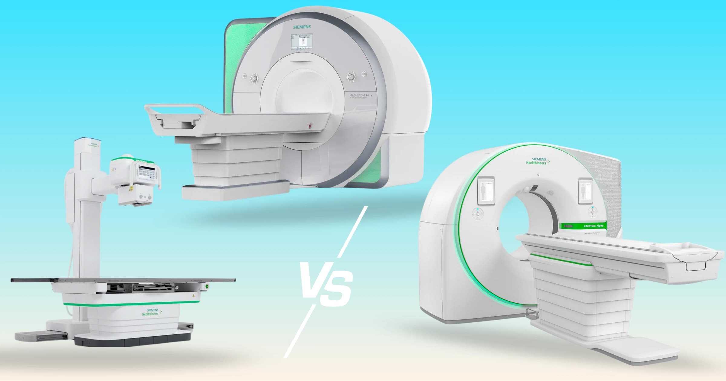
People sometimes notice that an X-ray technologist steps out of the room before pressing the button – and wonder, if it's safe for me, why do they leave the room?
The answer is that it's not about one scan being "unsafe" for you – it's about minimizing cumulative exposure for people who perform these scans hundreds or thousands of times a year.
Medical imaging exists on a spectrum of technologies – some use ionizing radiation, some don't, and each has different strengths.
Understanding the differences helps you make better, more confident choices.
Quick Takeaways
- MRI and ultrasound use no ionizing radiation – zero.
- X-rays and CT scans use ionizing radiation, but in doses that are generally safe when medically appropriate.
- Radiation dose is measured in millisieverts (mSv), and is best understood in the context of natural background radiation – the constant, low-level radiation we all receive from the sun, the ground, and everyday life, which averages about 3 mSv per year.
Radiation Dose by Modality
No Radiation
- MRI: Uses magnets and radiofrequency waves to create detailed images of soft tissue, joints, the brain, spine, and more.
- Ultrasound: Uses high-frequency sound waves; great for pregnancy, abdominal organs, and blood vessels.
Low Dose
- Chest X-ray: ~0.1 mSv (≈10 days of background radiation)
- Dental X-ray: ~0.004 mSv (≈1 day of background radiation)
- Mammogram: ~0.4 mSv (≈7 weeks of background radiation)
Moderate Dose
- CT Head: ~2 mSv (≈8 months of background radiation)
- CT Chest: ~7 mSv (≈2 years of background radiation)
- CT Abdomen/Pelvis: ~8 mSv (≈2.5 years of background radiation)
Higher Dose
- PET/CT: ~25 mSv (≈8 years of background radiation)
Perspective: Even at the higher end, the absolute risk from a single scan is very small – and if it finds or rules out something serious, the benefit greatly outweighs the risk.
Radiation Doses Are Getting Lower
Average radiation doses have decreased significantly in recent years across all modalities that use ionizing radiation.
Many of the dose figures you see online are population averages that include scans from older machines.
Modern CT, PET/CT, and angiography systems have advanced dose-reduction features like:
- Iterative reconstruction algorithms
- Automatic tube current modulation
- Faster detectors requiring less exposure
- ECG gating and tailored scan ranges for cardiac imaging
Example: In coronary CT angiography (CCTA), mean effective dose has dropped from ~7.15 mSv in 2010 to ~2.88 mSv in 2019 – a 2.5× reduction – largely due to newer scanner technology.
Why This Matters for Patients
Radiation dose varies by facility, scanner type, and protocol. If you're getting a scan, you can ask the facility:
- "What is the average radiation dose for this procedure here?"
- "Do you use dose-optimized protocols for my body size?"
They should be able to provide this – and it's a sign of a quality-focused imaging center if they can answer clearly.
Patient Experience
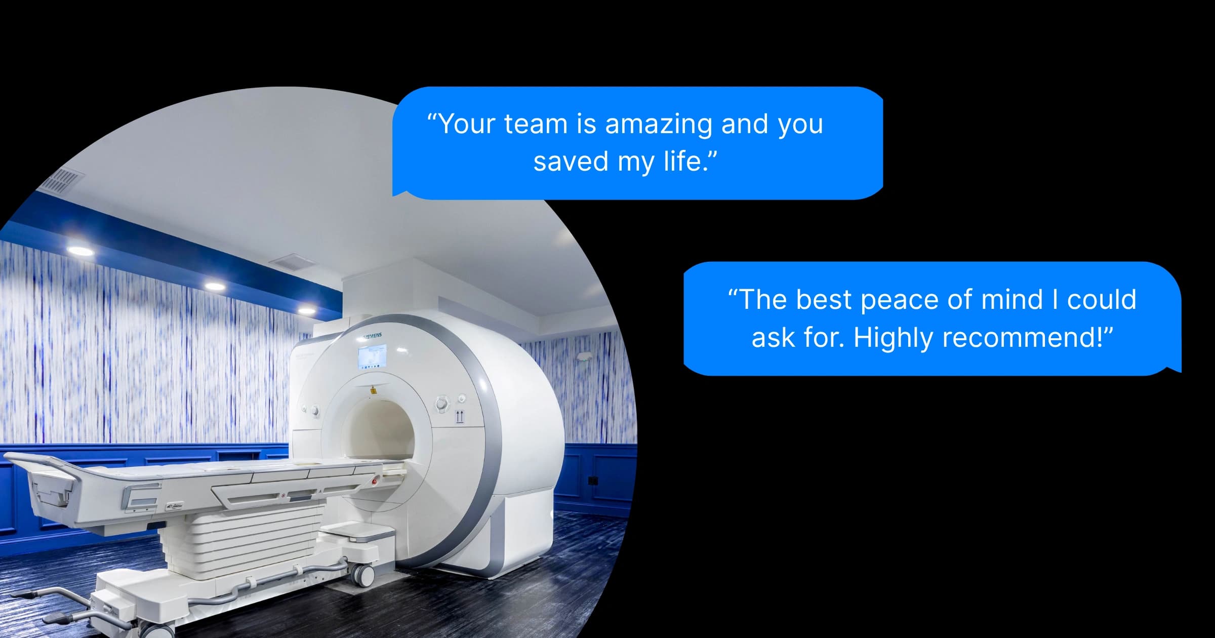
MRI – Takes 20–60 minutes. You'll lie on a narrow table that slides into a tube; newer wide-bore and open designs feel less confining. You'll hear loud tapping or knocking sounds, so earplugs or headphones are provided. It's important to remain still throughout the scan.
CT – Usually under 10 minutes. The scanner is a large, open "donut" shape. You lie on a table that moves through the opening. You might be asked to hold your breath briefly.
X-ray – Quick and simple, usually seconds to a few minutes. You'll be positioned in front of a detector or plate while a short exposure is taken.
Ultrasound – Takes 15–45 minutes. A technician applies gel to your skin and moves a handheld probe over the area. It's painless, quiet, and done while you're awake and alert.
When Each Modality is Chosen
| Modality | Strengths | Limitations | Typical Situations |
|---|---|---|---|
| MRI | Exceptional detail for soft tissues, nerves, and organs; no radiation; versatile imaging across body regions | Longer exam time (20–60 min); more expensive; confined space may cause discomfort | In-depth evaluation of brain, spine, joints, and soft tissue; follow-up scans without radiation concerns |
| CT | Very fast (often <10 min); excellent detail for bones, lungs, and internal organs; ideal for emergencies | Uses ionizing radiation (though significantly reduced with modern scanners); inferior soft tissue detail compared to MRI | Rapid assessment of trauma, stroke, chest or abdominal pain |
| X-ray | Quick (seconds to minutes); inexpensive; low radiation; good for initial bone and chest assessment | Limited detail for soft tissues; 2D projection may miss subtle issues | Detecting fractures, joint dislocations, lung infections, or dental problems |
| Ultrasound | No radiation; portable; real-time imaging; relatively low cost | Cannot penetrate bone or air; image quality varies with operator skill and patient anatomy | Pregnancy scans; abdominal organ evaluation; blood vessel studies; guiding needles or catheters |
Why Staff Step Out
Radiology staff limit their exposure to ionizing radiation over a career – even small doses add up when repeated thousands of times.
They rely on distance, lead shielding, and beam control. For a patient, occasional scans are well within safety limits.
Distance matters exponentially. Radiation intensity follows the inverse square law – step twice as far away, get four times less radiation.
Smart Imaging Choices
When your doctor recommends a scan, ask:
- What's the goal of this scan?
- Is there a radiation-free option that's equally effective for this case?
- How will this scan change my care?
- Do you have my prior imaging?
Keeping your own record of scans prevents unnecessary repeats – which can happen simply because records aren't shared between facilities.
The Bottom Line
- MRI and ultrasound: No radiation, excellent for many conditions, especially when repeat imaging is needed.
- CT and X-ray: Use radiation, but doses are much lower on modern scanners, and are safe when medically justified.
- PET/CT: Higher radiation doses, but unmatched for certain cancer staging and follow-up – and doses are trending downward here too.
The safest scan is the one that answers the clinical question with the least risk – and all modern imaging technologies have an important place in medicine.
Fascinating Further Readings
References
[1] WIS Admin. "Understanding Radiation From Mammograms – Are They Safe?" Accessed August 11, 2025. link
[2] The American Cancer Society medical and editorial content team. "Understanding Radiation Risk from Imaging Tests." Accessed August 11, 2025. link
[3] U.S. Environmental Protection Agency. "Frequent Questions about Radiation in Medicine." Accessed August 11, 2025. link
[4] Stanford University Environmental Health & Safety. "Lead Apron Use Policy." Accessed August 11, 2025. link
[5] Linet, Martha S., et al. "Exposure of U.S. Populations to Radiofrequency Electromagnetic Fields from Medical Procedures." PLoS Medicine 13, no. 8 (2016): e1002192. link
[6] Masoumeh Karimi, et al. "Diagnostic Radiation Exposure and Cancer Risk." International Journal of Cancer Management 14, no. 1 (2021): e161617. link
[7] Shawna Seed. "Radiation Doses in CT Scans." Accessed August 11, 2025. link
[8] "How Much Radiation Do You Get from Imaging Tests?" Accessed August 11, 2025. link
[9] "Comparison of radiation dose and its correlates between coronary computed tomography angiography and invasive coronary angiography in Northeastern Thailand." Accessed August 13, 2025. link


